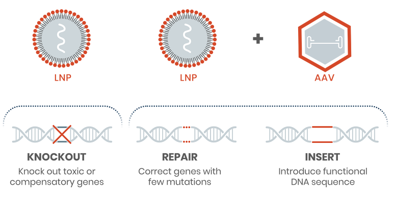
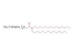
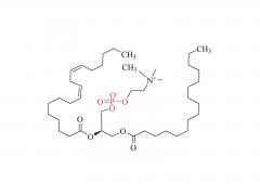
Industry News
  • PEG hydrogel for in vivo hemostasis
    PEG hydrogel for in vivo hemostasis May 14,2020.
    A sealant can significantly improve the effect of visceral surgery; it can not only reduce intraoperative blood loss, but also reduce postoperative complications such as secondary hemorrhage and tissue adhesion, which are essential in surgical operations. However, the sealant currently used for in vivo hemostasis cannot address the needs in the modern aging society. The main challenges are its safeness, easiness of preparation and removal, and price. The commercial synthetic sealants are mainly made up of PEG, for example the 4-arm PEG hydrogel based on the ammonolysis reaction. Those sealants have advantages of high strength, strong adhesion and economic price, but the disadvantage is that they cannot be quickly degraded and can easily cause foreign body reaction in the wound that leads to healing delay. In order to overcome the limitations of the existing PEG hydrogels, a new PEG sealant based on multi-arm PEG Succinimidyl Succinate (amide bond) has been jointly developed by Institute of Chemistry, Chinese Academy of Science and the General Hospital of People's Liberation Army. The in vitro experiments show that SS glue has a better hemostatic effect than the previously developed SG and gauze. SS can quickly stanch the bleeding on the wound as well as prevent the adhesion issue after the operation. In contrast, SG and gauze both have different degree of postoperative adhesion when they are used for hemostasis. However, this is not the case for SS, as it is able to stop bleeding effectively even for patients taking anticoagulants, which cannot be achieved by the widely used fibrin glue. The researchers compare the hemostatic effects of SS, SG and gauze on wounds. Among them, SS and SG can achieve rapid wound hemostasis, while gauze is much slower. And after a week of hemostasis, both SG and gauze have different degrees of adhesion while SS does not have such side effects. It indicates that SS not only can stop bleeding, but also acts as a physical barrier to prevent the wound from adhering to the surrounding tissues during the healing process (Figure a). Figure b compares the healing situation of wounds at different times after surgery. Figure c compares the separate hemostatic effects of SS and fibrin glue used in the wounds of a New Zealand white rabbit with anticoagulants. SS has a better hemostatic effect than fibrin glue in terms of speed and stability. The author further uses SS to perform the hemostasis experiment on a large wound surface (diameter: 25mm, depth: 10mm). Even if a coagulant is used, SS can effectively stop bleeding after a certain period of time. [1] Bu Y ,  Zhang L ,  Sun G , et al. Tetra㏄EG Based Hydrogel Sealants for In Vivo Visceral Hemostasis[J]. Advanced Materials, 2019, 31(28):1901580.1-1901...
    View More
  • Amyloid-Like Rapid Surface Modification for Antifouling and In-Depth Remineralization of Dentine Tubules to Treat Dental Hypersensitivity
    Amyloid-Like Rapid Surface Modification for Antifouling and In-Depth Remineralization of Dentine Tubules to Treat Dental Hypersensitivity January 6,2020.
    Exposure of dentinal tubules (DTs) leads to the transmission of external stimuli within the DTs, causing dental hypersensitivity (DH). Approximately 42% 18- to 35-year-olds experience dental hypersensitivity (DH), which is characterized by a short or transient sharp pain arising from exposed dentin. To treat DH, various desensitizers have been developed for occluding DTs. However, most desensitizers commercially available or in development are only able to seal the orifices, rather than the deep regions of the DTs, thus lacking long-term stability. Dr. C. Li, Prof. P. Yang found it is shown that the fast amyloid-like aggregation of lysozyme (lyso) conjugated with poly(ethylene glycol) (PEG) (lyso-PEG) can afford a robust ultrathin nanofilm on the deep walls of DTs through a rapid one-step aqueous coating process (in 2 min). The resultant nanofilm provides a highly effective antifouling platform for resisting the attachment of oral bacteria such as Streptococcus mutans and induces remineralization in the DTs to seal both the orifices and depths of the DTs by forming hydroxyapatite (HAp) minerals in situ. Both in vitro and in vivo animal experiments prove that the nanofilm-coated DTs are occluded with a depth of over 60 ± 5 µm, which is at least 6 times deeper than that reported in the literature. This approach thus demonstrates the concept that an amyloid-like proteinaceous nanofilm can offer an inexpensive, rapid, and efficient therapy for treating DH with long-term effect. Sinopeg provide various NW poly(ethylene glycol) (PEG) products: 2KDa, 5KDa, 10KDa, 20KDa, etc. Products: Linear Monofunctional PEGs Linear Bifunctional PEGs Linear Heterofunctional PEGs Branched PEGs Multi-Arm Functional PEGs Functionally Grafted PEGs
    View More
  • Tetra-PEG Based Hydrogel Sealants for In Vivo Visceral  Hemostasis
    Tetra-PEG Based Hydrogel Sealants for In Vivo Visceral Hemostasis 2019-11-08
    Tetra-PEG hydrogels based on the ammonolysis reaction between tetra-armed poly(ethylene glycol) amine (Tetra-PEG-NH2) and Tetra-PEG-SAE offer massive advantages as sealants. They are entirely synthetic without the misgivings of being inhibited by anticoagulation agents and transferring disease. Their cost is low due to their easily preservable components with high accessibility. Because of the intrinsic properties of this ammonolysis reaction, the resulting hydrogels can gel  fast just by injection and adhere to the tissues tightly throughchemical bonds. Another remarkable advantage for Tetra-PEG hydrogels is that they are mechanically tough, and the sealants are favored to be mechanically tough to keep stable in case of dynamic movement of the tissues and the use of assistant pressure which is a key adjunctive step in achieving hemostasis. However, two hurdles are preventing extending their applications in vivo. The first one is that just as commercialized sealants, none of the reported Tetra-PEG hydrogels could be controllably removed without mechanical debridement, which is extremely dangerous because of their high mechanical strength. Besides, they possess long degradation time, which will lead to severe foreign body reactions, tissue adhesion, disturbed tissue healing, and obstruction of the circulatory system, when used in vivo. Here, to overcome the limitations of the existing ammonolysis based Tetra-PEG hydrogels, we construct an optimized one (SS) with fast degradable and controllably dissolvable properties via Tetra-PEG-NH2 and tetra-armed poly(ethylene glycol) succinimidyl succinate (Tetra-PEG-SS) . The resulting SS exhibits biocompatibility superior to the reported degradable Tetra-PEG hydrogel (SG) based on Tetra-PEG-NH2 and tetra-armed poly(ethylene glycol) succinimidyl glutarate (Tetra-PEG-SG) . More importantly, in contrast to the disappointing results of SG that leads to serious adverse effects in in vivo hemostasis due to the long retention, SS causes almost no noticeable side effects with outstanding hemostasis efficacy even under the anticoagulated situations. This hydrogel is a promising candidate for the next-generation in vivo sealants in the aged society.
    View More
  • Lipid Nanoparticle Systems for Enabling Gene Therapies
    Lipid Nanoparticle Systems for Enabling Gene Therapies August 11,2025.
    Review Mol Ther. 2017 Jul 5;25(7):1467-1475.  doi: 10.1016/j.ymthe.2017.03.013.  Epub 2017 Apr 13. Lipid Nanoparticle Systems for Enabling Gene Therapies Abstract Genetic drugs such as small interfering RNA (siRNA), mRNA, or plasmid DNA provide potential gene therapies to treat most diseases by silencing pathological genes, expressing therapeutic proteins, or through gene-editing applications.  In order for genetic drugs to be used clinically, however, sophisticated delivery systems are required.  Lipid nanoparticle (LNP) systems are currently the lead non-viral delivery systems for enabling the clinical potential of genetic drugs.  Application will be made to the Food and Drug Administration (FDA) in 2017 for approval of an LNP siRNA drug to treat transthyretin-induced amyloidosis, presently an untreatable disease.  Here, we first review research leading to the development of LNP siRNA systems capable of silencing target genes in hepatocytes following systemic administration.  Subsequently, progress made to extend LNP technology to mRNA and plasmids for protein replacement, vaccine, and gene-editing applications is summarized.  Finally, we address current limitations of LNP technology as applied to genetic drugs and ways in which such limitations may be overcome.  It is concluded that LNP technology, by virtue of robust and efficient formulation processes, as well as advantages in potency, payload, and design flexibility, will be a dominant non-viral technology to enable the enormous potential of gene therapy. Keywords: gene editing;  gene therapy;  genetic drugs;  lipid nanoparticles;  mRNA;  siRNA. For more product information, please contact us at: US Tel: 1-844-782-5734 US Tel: 1-844-QUAL-PEG CHN Tel: 400-918-9898 Email: sales@sinopeg.com
    View More
first page 21 22 23 last page

A total of 23 pages

Copyright © XIAMEN SINOPEG BIOTECH CO., LTD. All Rights Reserved.

Home

Products

News

contact