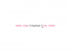
Industry News
  • PEG hydrogel for in vivo hemostasis
    PEG hydrogel for in vivo hemostasis May 14,2020.
    A sealant can significantly improve the effect of visceral surgery; it can not only reduce intraoperative blood loss, but also reduce postoperative complications such as secondary hemorrhage and tissue adhesion, which are essential in surgical operations. However, the sealant currently used for in vivo hemostasis cannot address the needs in the modern aging society. The main challenges are its safeness, easiness of preparation and removal, and price. The commercial synthetic sealants are mainly made up of PEG, for example the 4-arm PEG hydrogel based on the ammonolysis reaction. Those sealants have advantages of high strength, strong adhesion and economic price, but the disadvantage is that they cannot be quickly degraded and can easily cause foreign body reaction in the wound that leads to healing delay. In order to overcome the limitations of the existing PEG hydrogels, a new PEG sealant based on multi-arm PEG Succinimidyl Succinate (amide bond) has been jointly developed by Institute of Chemistry, Chinese Academy of Science and the General Hospital of People's Liberation Army. The in vitro experiments show that SS glue has a better hemostatic effect than the previously developed SG and gauze. SS can quickly stanch the bleeding on the wound as well as prevent the adhesion issue after the operation. In contrast, SG and gauze both have different degree of postoperative adhesion when they are used for hemostasis. However, this is not the case for SS, as it is able to stop bleeding effectively even for patients taking anticoagulants, which cannot be achieved by the widely used fibrin glue. The researchers compare the hemostatic effects of SS, SG and gauze on wounds. Among them, SS and SG can achieve rapid wound hemostasis, while gauze is much slower. And after a week of hemostasis, both SG and gauze have different degrees of adhesion while SS does not have such side effects. It indicates that SS not only can stop bleeding, but also acts as a physical barrier to prevent the wound from adhering to the surrounding tissues during the healing process (Figure a). Figure b compares the healing situation of wounds at different times after surgery. Figure c compares the separate hemostatic effects of SS and fibrin glue used in the wounds of a New Zealand white rabbit with anticoagulants. SS has a better hemostatic effect than fibrin glue in terms of speed and stability. The author further uses SS to perform the hemostasis experiment on a large wound surface (diameter: 25mm, depth: 10mm). Even if a coagulant is used, SS can effectively stop bleeding after a certain period of time. [1] Bu Y ,  Zhang L ,  Sun G , et al. Tetra㏄EG Based Hydrogel Sealants for In Vivo Visceral Hemostasis[J]. Advanced Materials, 2019, 31(28):1901580.1-1901...
    View More
  • Amyloid-Like Rapid Surface Modification for Antifouling and In-Depth Remineralization of Dentine Tubules to Treat Dental Hypersensitivity
    Amyloid-Like Rapid Surface Modification for Antifouling and In-Depth Remineralization of Dentine Tubules to Treat Dental Hypersensitivity January 6,2020.
    Exposure of dentinal tubules (DTs) leads to the transmission of external stimuli within the DTs, causing dental hypersensitivity (DH). Approximately 42% 18- to 35-year-olds experience dental hypersensitivity (DH), which is characterized by a short or transient sharp pain arising from exposed dentin. To treat DH, various desensitizers have been developed for occluding DTs. However, most desensitizers commercially available or in development are only able to seal the orifices, rather than the deep regions of the DTs, thus lacking long-term stability. Dr. C. Li, Prof. P. Yang found it is shown that the fast amyloid-like aggregation of lysozyme (lyso) conjugated with poly(ethylene glycol) (PEG) (lyso-PEG) can afford a robust ultrathin nanofilm on the deep walls of DTs through a rapid one-step aqueous coating process (in 2 min). The resultant nanofilm provides a highly effective antifouling platform for resisting the attachment of oral bacteria such as Streptococcus mutans and induces remineralization in the DTs to seal both the orifices and depths of the DTs by forming hydroxyapatite (HAp) minerals in situ. Both in vitro and in vivo animal experiments prove that the nanofilm-coated DTs are occluded with a depth of over 60 ± 5 µm, which is at least 6 times deeper than that reported in the literature. This approach thus demonstrates the concept that an amyloid-like proteinaceous nanofilm can offer an inexpensive, rapid, and efficient therapy for treating DH with long-term effect. Sinopeg provide various NW poly(ethylene glycol) (PEG) products: 2KDa, 5KDa, 10KDa, 20KDa, etc. Products: Linear Monofunctional PEGs Linear Bifunctional PEGs Linear Heterofunctional PEGs Branched PEGs Multi-Arm Functional PEGs Functionally Grafted PEGs
    View More
  • Tetra-PEG Based Hydrogel Sealants for In Vivo Visceral  Hemostasis
    Tetra-PEG Based Hydrogel Sealants for In Vivo Visceral Hemostasis 2019-11-08
    Tetra-PEG hydrogels based on the ammonolysis reaction between tetra-armed poly(ethylene glycol) amine (Tetra-PEG-NH2) and Tetra-PEG-SAE offer massive advantages as sealants. They are entirely synthetic without the misgivings of being inhibited by anticoagulation agents and transferring disease. Their cost is low due to their easily preservable components with high accessibility. Because of the intrinsic properties of this ammonolysis reaction, the resulting hydrogels can gel  fast just by injection and adhere to the tissues tightly throughchemical bonds. Another remarkable advantage for Tetra-PEG hydrogels is that they are mechanically tough, and the sealants are favored to be mechanically tough to keep stable in case of dynamic movement of the tissues and the use of assistant pressure which is a key adjunctive step in achieving hemostasis. However, two hurdles are preventing extending their applications in vivo. The first one is that just as commercialized sealants, none of the reported Tetra-PEG hydrogels could be controllably removed without mechanical debridement, which is extremely dangerous because of their high mechanical strength. Besides, they possess long degradation time, which will lead to severe foreign body reactions, tissue adhesion, disturbed tissue healing, and obstruction of the circulatory system, when used in vivo. Here, to overcome the limitations of the existing ammonolysis based Tetra-PEG hydrogels, we construct an optimized one (SS) with fast degradable and controllably dissolvable properties via Tetra-PEG-NH2 and tetra-armed poly(ethylene glycol) succinimidyl succinate (Tetra-PEG-SS) . The resulting SS exhibits biocompatibility superior to the reported degradable Tetra-PEG hydrogel (SG) based on Tetra-PEG-NH2 and tetra-armed poly(ethylene glycol) succinimidyl glutarate (Tetra-PEG-SG) . More importantly, in contrast to the disappointing results of SG that leads to serious adverse effects in in vivo hemostasis due to the long retention, SS causes almost no noticeable side effects with outstanding hemostasis efficacy even under the anticoagulated situations. This hydrogel is a promising candidate for the next-generation in vivo sealants in the aged society.
    View More
  • Safety profile of two-dimensional Pd nanosheets for photothermal therapy and photoacoustic imaging
    Safety profile of two-dimensional Pd nanosheets for photothermal therapy and photoacoustic imaging January 18,2024.
    Research Article | Issue | Published: 06 December 2016 Safety profile of two-dimensional Pd nanosheets for photothermal therapy and photoacoustic imaging Mei Chen1,§, Shuzhen Chen2,3,§, Chengyong He2,§, Shiguang Mo1, Xiaoyong Wang2, Gang Liu2, Nanfeng Zheng1 Abstract Two-dimensional (2D) nanosheets have emerged as an important class of nanomaterial with great potential in the field of biomedicines, particularly in cancer theranostics.   However, owing to the lack of effective methods that synthesize uniform 2D nanomaterials with controlled size, systematic evaluation of size-dependent bio-behaviors of 2D nanomaterials is rarely reported.   To the best of our knowledge, we are the first to report a systematic evaluation of the influence of size of 2D nanomaterials on their bio-behaviors.   2D Pd nanosheets with diameters ranging from 5 to 80 nm were synthesized and tested in cell and animal models to assess their size-dependent bioapplication, biodistribution, elimination, toxicity, and genomic gene expression profiles.   Our results showed size significantly influences the biological behaviors of Pd nanosheets, including their photothermal and photoacoustic effects, pharmacokinetics, and toxicity.   Compared to larger-sized Pd nanosheets, smaller-sized Pd nanosheets exhibited more advanced photoacoustic imaging and photothermal effects upon ultralow laser irradiation.   Moreover, in vivo results indicated that 5-nm Pd nanosheets escape from the reticuloendothelial system with a longer blood half-life and can be cleared by renal excretion, while Pd nanosheets with larger sizes mainly accumulate in the liver and spleen.   The 30-nm Pd nanosheets exhibited the highest tumor accumulation.   Although Pd nanosheets did not cause any appreciable toxicity at the cellular level, we observed slight lipid accumulation in the liver and inflammation in the spleen.   Genomic gene expression analysis showed that 80-nm Pd nanosheets interacted with more cellular components and affected more biological processes in the liver, as compared to 5-nm Pd nanosheets.   We believe this work will provide valuable information and insights into the clinical application of 2D Pd nanosheets as nanomedicines. Related products Abbreviation: mPEG-SH Name: Methoxypoly(ethylene glycol) thiol For more product information, please contact us at: US Tel: 1-844-782-5734 US Tel: 1-844-QUAL-PEG CHN Tel: 400-918-9898 Email: sales@sinopeg.com
    View More
  • Engineering PEG-based hydrogels to foster efficient endothelial network formation in free-swelling and confined microenvironments
    Engineering PEG-based hydrogels to foster efficient endothelial network formation in free-swelling and confined microenvironments June 8,2020.
    Engineering PEG-based hydrogels can efficiently foster endothelial network formation in free-swelling and confined microenvironments Polyethylene glycol (PEG) and its derivatives are among the few polymers approved by the US FDA that can be used in biomedical products. The PEGl-based hydrogel has excellent flexibility and biocompatibility. Some PEG hydrogels can not only be degraded, but also can form bioactive site through modifying the connexins in a chemical way. In vitro tissue engineered models are expected to have significant impact on disease modeling and preclinical drug development. Reliable methods to induce microvascular networks in such microphysiological systems are needed to improve the size and physiological function of these models. By systematically engineering several physical and biomolecular properties of the cellular microenvironment (including crosslinking density, polymer density, adhesion ligand concentration, and degradability), the author Alexander Brown establish design principles that describe how synthetic matrix properties influence vascular morphogenesis in modular and tunable hydrogels based on commercial 8-arm poly (ethylene glycol) (PEG8a) macromers. The author applies these design principles to generate endothelial networks that exhibit consistent morphology throughout depths of hydrogel greater than 1 mm. These PEG8a-based hydrogels have relatively high volumetric swelling ratios (>1.5), which limits their utility in confined environments such as microfluidic devices. To overcome this limitation, the author mitigates swelling by incorporating a highly functional PEG-grafted alpha-helical poly (propargyl-l-glutamate) (PPLGgPEG) macromer along with the canonical 8-arm PEG8a macromer in gel formation. This hydrogel platform supports enhanced endothelial morphogenesis in neutral-swelling environments. Finally, the author incorporates PEG8a-PPLGgPEG gels into microfluidic devices and demonstrates improved diffusion kinetics and microvascular network formation in situ compared to PEG8a-based gels. [1] Brown A ,  He H ,  Trumper E , et al. Engineering PEG-based hydrogels to foster efficient endothelial network formation in free-swelling and confined microenvironments[J]. Biomaterials, 2020, 243:119921. If there is any copyright infringement, please contact us and we will remove the content at the first time. Sinopeg provide various NW poly(ethylene glycol) (PEG) products: 2KDa, 5KDa, 10KDa, 20KDa, etc. Products: Linear Monofunctional PEGs Linear Bifunctional PEGs Linear Heterofunctional PEGs Branched PEGs Multi-Arm Functional PEGs Functionally Grafted PEGs
    View More
first page 11 12 13 14 last page

A total of 14 pages

Copyright © XIAMEN SINOPEG BIOTECH CO., LTD. All Rights Reserved.

Home

Products

News

contact